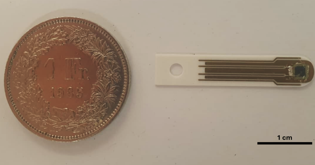
Free-standing SiC membranes provide an effective solution for non-invasive X-ray beam monitoring, particularly at hard and tender X-ray energies. However, a transmission-type detector is not always compatible with the experimental conditions.
At soft X-ray energies, even extremely thin SiC membranes can introduce non-negligible absorption, reducing the photon flux delivered to the sample. In these cases, beam diagnostics must be implemented without placing additional material directly in the useful X-ray beam.
Turning existing optical components into active beam monitors, functionalizing X-ray optics
In scanning transmission X-ray microscopy (STXM), a Fresnel Zone Plate focuses the X-ray beam onto the sample. Since only a fraction of the incident radiation contributes to the useful focused beam, additional optical elements such as the Center Stop (CS) and Order-Sorting Aperture (OSA) are used to suppress the unfocused 0ᵗʰ order and higher diffraction orders.
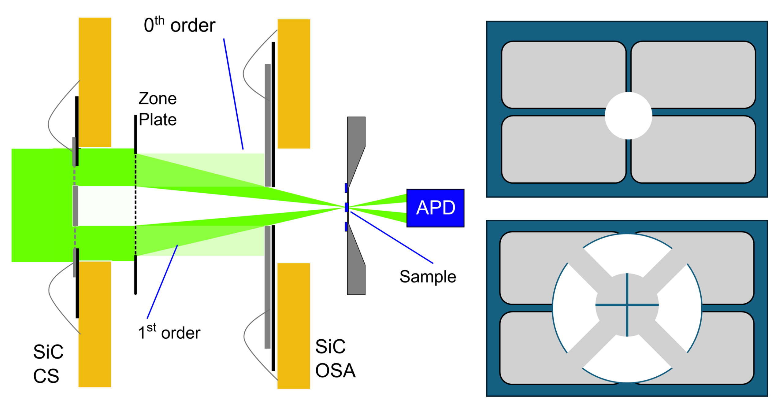
Instead of simply blocking this otherwise unused radiation, we developed a different concept:
make these optical elements radiation-sensitive.
By fabricating the OSA and Center Stop directly from active SiC diode structures, the same component can perform both an optical function and a beam-diagnostic function.
What can be measured?
The SiC optical elements can be segmented into multiple electrically independent regions, allowing the photocurrent generated by the X-ray beam to provide information on:
• Incident beam intensity, I₀
• Transverse beam position and displacement
• Beam stability during image or spectrum acquisition
The approach therefore enables continuous, in-situ beam monitoring at a position very close to the experimental conditions experienced by the sample, rather than relying only on diagnostics located further upstream or on separate reference measurements.
For spectromicroscopy applications, access to a simultaneous I₀ signal can also support improved normalization of the acquired data and, potentially, correction of beam-intensity fluctuations during acquisition.
From concept to beamline validation
SiC-based OSA and Center Stop devices have been fabricated and experimentally tested at the PolLux STXM beamline of the Swiss Light Source. Installation required only minor modifications to the existing endstation, mainly the addition of the electrical connections needed for detector readout.
During the experimental validation, the functionalized OSA simultaneously detected beam-intensity and position variations. Between 700 and 1000 eV, the SiC signal showed a strong linear correlation with the conventional STXM reference detector, with R² = 0.997.
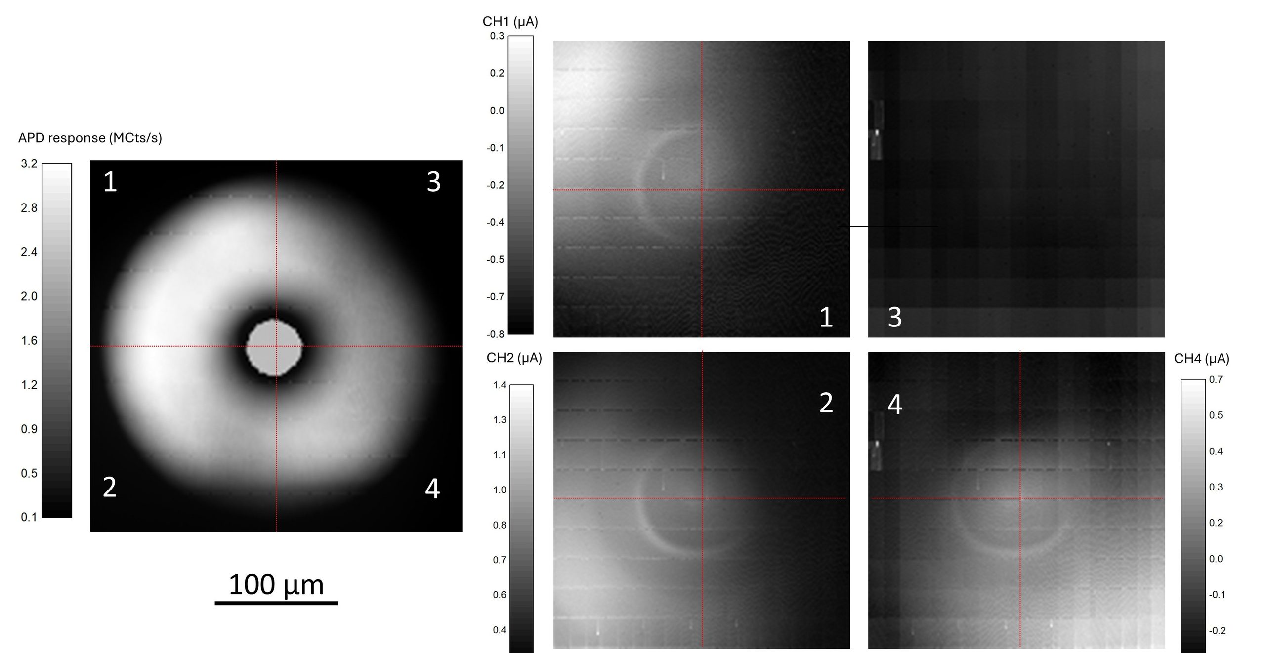
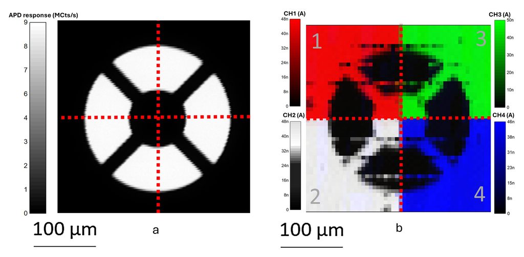
These results demonstrate that SiC detectors can be integrated directly into X-ray optical components while preserving both their optical function and their capability as active radiation sensors.
Interested in integrating beam diagnostics into an existing optical component?
Contact us to discuss feasibility and application-specific development.
Publications about this technology
| G. Trovato, S. Finizio, S. Moscato, J. Raabe, J. Reuteler and M. Camarda, Monolithic integration of SiC diode detectors into scanning X-ray microscopy optical elements for in situ beam diagnostics, Journal of Synchrotron Radiation, 33, 2026. |